In MS-like mice, immune cells increased near smell nerves
By MS Buddy ·
Summarized from Science advances, Jun 2025 · PubMed · DOI. AI-generated summary. Not medical advice.
Animal study · mice with an MS-like disease or a brain infection
Research by Collin Laaker, Zsuzsanna Fabry and 10 colleagues at the University of Wisconsin-Madison in the United States.
How do immune cells near smell nerves change in mice with an MS-like disease?
Key Takeaway
In mice with an MS-like disease, immune cells increased around nerves used for smell, and nearby skull bone marrow changed.
What They Found
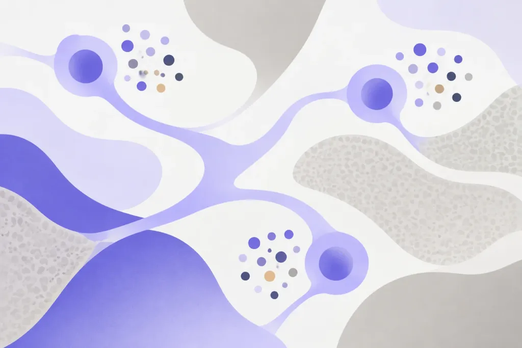
Researchers studied mice with an MS-like disease and mice with a brain infection. During disease, more immune cells gathered around nerves used for smell. Material placed inside the skull reached these cells. Small channels connected the area around those nerves to bone marrow in the skull. In mice with the MS-like disease, that bone marrow produced more immune cells and received material draining from inside the skull.
Who Should Care and Why
This study examined mice, not people with MS. It focused on the nerves used for smell and nearby skull bone marrow. These areas connect to a route that drains fluid from around the brain. The findings show how immune activity changed along that route in mice with an MS-like disease. Mice with a brain infection also showed that bacteria could reach the nearby bone marrow.
Important Considerations
You cannot assume the same changes happen in people with MS. The abstract does not give the number of mice studied. This study did not test a treatment or measure whether these changes affected symptoms.
AI-generated summary — for informational purposes only, not medical advice
Read the original abstract
Published in Science advances by et al.. Reproduced from the PubMed record.
Cerebrospinal fluid (CSF) contains inflammatory cues that enable peripheral immune surveillance of the central nervous system (CNS). While some cranial nerves allow for CSF efflux, the immune environment around CSF-interfacing cranial nerves during neuroinflammation is still poorly understood. Using a mouse model of multiple sclerosis [experimental autoimmune encephalomyelitis (EAE)] and CNS infection (CNS-Mtb), we examined immune responses around olfactory nerve bundles near the cribriform plate, a key CSF efflux route. During neuroinflammation, we found increased perineural immune cells that had access to intracranial injected beads, dye, and bacteria. Additionally, we identified osseous channels connecting the environment surrounding olfactory nerves to bone marrow in the cribriform plate (cpBM). Notably, the cpBM undergoes myelopoiesis during EAE, has access to components of intracranial drainage, and is vulnerable to Mtb bacteria invasion during CNS-Mtb infection. Our findings improve the understanding of how the environments of CSF-interfacing cranial nerves and bone marrow are altered within the skull during neuroinflammatory disease.
Categories
Explore more research
Check out the MS Buddy app
It’s free, with no ads. Built by people living with MS.


Related MS research
11/15/2026
In tissue from mice with an MS-like disease, a lab drug reduced excess chemical release from nerve e
Read More
11/13/2026
A review compares MS with harmful drug reactions and proposes ways to test how immune genes, lasting
Read More
11/13/2026
This review examines how vitamin D and vitamin A signals shape immune activity in MS and other disea
Read More
11/12/2026
In mice with an MS-like disease, a lab-made protein improved movement and thinking. Researchers also
Read More
11/12/2026
A review proposes how the Epstein-Barr virus might contribute to MS. The model brings earlier ideas
Read More
11/11/2026
This review compares inherited and environmental risks for MS starting in childhood or adulthood, in
Read MoreNot all studies carry the same weight: sample size, study design and whether results were replicated all matter. The credibility score above reflects the journal, and the original paper is linked under More Details. Read how MS Buddy selects and scores research, and talk to your MS care team before acting on any study.