Lining up MS brain scans gave stronger scan readings
By MS Buddy ·
Summarized from Magnetic resonance imaging, Nov 2026 · PubMed. AI-generated summary. Not medical advice.
Observational study · 26 people with MS who had brain scans at different angles
Research by Muhammed Zayed, Amir Eissa and 3 colleagues at Al-Azhar University and Suez Canal University in Egypt.
Does the angle of a brain scan change its readings in people with MS?
Key Takeaway
In people with MS, brain scan readings were stronger when the images lined up with the scanner’s magnetic field.
What They Found
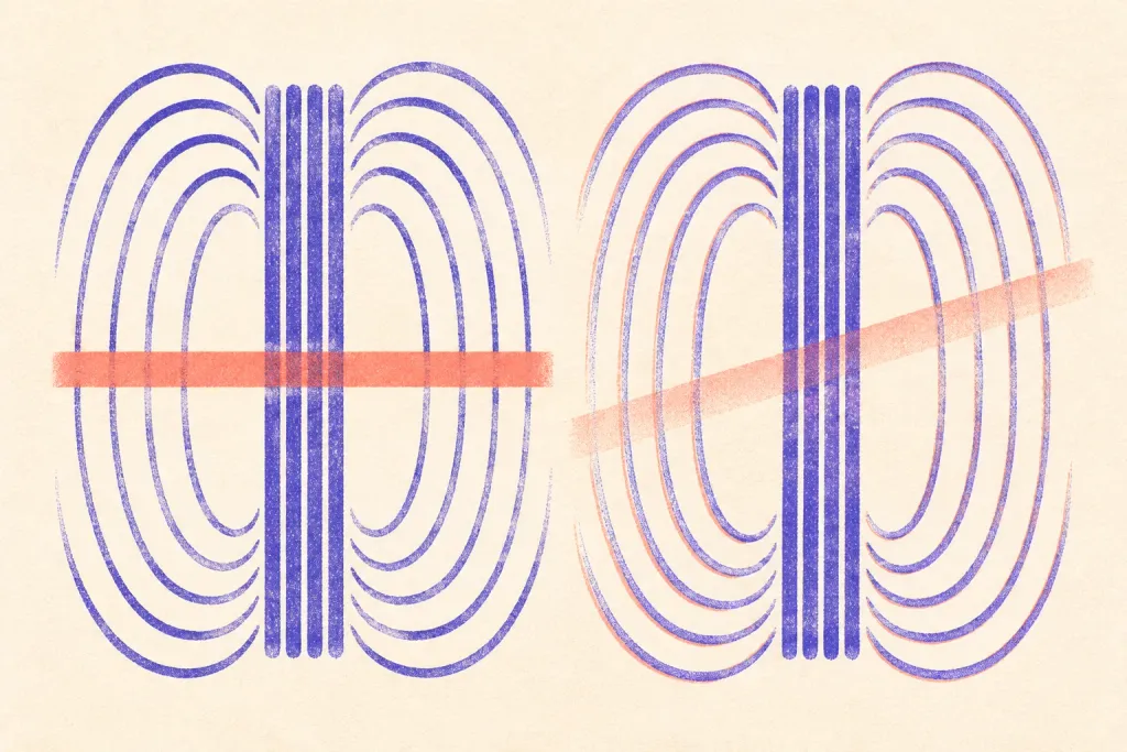
Each person had two brain scans using different image angles. One scan lined up with the scanner’s magnetic field. The other lined up with landmarks in the brain. Readings were stronger with the first approach across all seven areas studied on both sides of the brain. The readings grew weaker as the image angle moved farther from the magnetic field.
Who Should Care and Why
The study included people with MS. It focused on areas deep inside the brain. The scans can pick up signs of iron buildup and small changes in brain tissue. The findings show that the angle of the scan affects how strongly those signs show up. The researchers suggest using the same angle to make readings easier to compare over time.
Important Considerations
Researchers planned the study in advance and compared two scan angles in the same 26 people. They used one scanner. The study did not test whether changing the scan angle helps track MS worsening or treatment results over time.
AI-generated summary — for informational purposes only, not medical advice
Read the original abstract
Published in Magnetic resonance imaging by et al.. Reproduced from the PubMed record.
Phase quantification has emerged as a critical method for monitoring disease progression and evaluating treatment efficacy in Multiple Sclerosis (MS) due to its high sensitivity to pathological iron deposition and microstructural changes. Despite its utility, most researchers continue to prioritize anatomical alignment protocols such as orientation parallel to the anterior-posterior commissure AC-PC line, which often result in slice angulations that deviate significantly, sometimes reaching 20ο, from the main magnetic field (B0). This prospective study evaluated the impact of such orientation variations on Susceptibility-Weighted Angiography (SWAN) phase values in twenty-six MS patients using a 1.5 T scanner. Each subject underwent two SWAN acquisitions: a standardized zero-alignment to B0 and a conventional anatomical alignment. Quantitative ROI analysis across seven bilateral deep gray matter structures revealed that phase magnitudes were consistently and significantly higher under zero alignment compared to anatomical alignment (p < 0.05). Furthermore, a progressive reduction in phase magnitude was observed as the angular deviation from B0 increased, confirming a clear inverse relationship between acquisition obliquity and susceptibility sensitivity. These findings suggest that current anatomical alignment practices may sub-optimize the detection of susceptibility-related biomarkers. Consequently, standardizing acquisitions to zero alignment is essential for ensuring the reproducibility of phase quantification and enhancing its clinical value for monitoring disease and therapeutic outcomes in MS.
Categories
Explore more research
Check out the MS Buddy app
It’s free, with no ads. Built by people living with MS.


Related MS research

11/20/2026
In a study of 140 people, a spinal fluid test better separated MS from other nervous system conditio
Read More
11/20/2026
A study of 59 people with relapsing-remitting MS found a computer tool helped doctors read brain sca
Read More
11/19/2026
A Japanese hospital study linked MS to about 9% of first eye nerve inflammation cases. Other immune
Read More
11/18/2026
An adjusted MRI scan found more veins within MS brain lesions in 17 people with MS. It improved scan
Read More
1/1/2026
After a car crash, brain and spine scans revealed signs of MS in an 18-year-old male. Further tests
Read More
11/14/2026
An opinion paper explains why MS brain scans can show tissue loss without proving nerve signal failu
Read MoreNot all studies carry the same weight: sample size, study design and whether results were replicated all matter. The credibility score above reflects the journal, and the original paper is linked under More Details. Read how MS Buddy selects and scores research, and talk to your MS care team before acting on any study.