Adjusted MRI spots more veins inside MS brain lesions
By MS Buddy ·
Summarized from European radiology, Nov 2026 · PubMed · DOI. AI-generated summary. Not medical advice.
Observational study · 17 people with a clinical diagnosis of MS
Research by Yin-Chen Hsu, Pascal Sati and 15 colleagues at Cedars-Sinai Medical Center, Biomedical Imaging Research Institute and 1 other institution in the United States.
Can an adjusted MRI scan detect more veins inside MS brain lesions?
Key Takeaway
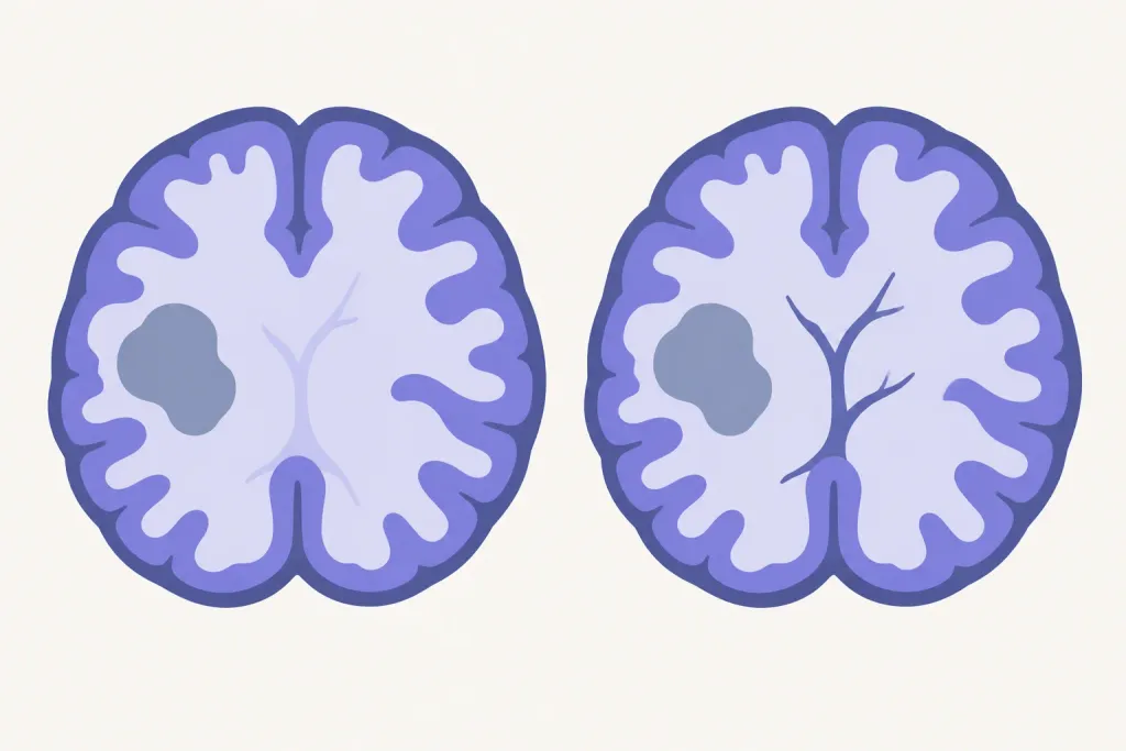
What They Found
The adjusted scan found 105 brain lesions (damaged areas) with a visible central vein, compared with 72 using the standard scan. It found every vein-containing lesion seen on the standard scan, plus 33 more. The adjusted scan found more vein-containing lesions in 14 of 17 participants. Scan readers were more confident in identifying these veins with the adjusted scan. Using a scan-based rule, positive results for MS rose from about 41% to 82%, without increasing scan time.
Who Should Care and Why
The study involved people already diagnosed with MS. It examined a scan feature that can help distinguish MS damage from similar-looking damage caused by other conditions. That feature is a vein running through the center of a brain lesion (a damaged area). The study focused on detecting this feature more clearly, rather than treating MS. The findings concern how scans are collected and read during MS assessment.
Important Considerations
This small study compared standard and adjusted scans in 17 people with MS. Two scan specialists assessed the images without knowing which scan method produced them. Because all participants already had MS, the study does not show how well the adjusted scan distinguishes MS from other conditions.
AI-generated summary — for informational purposes only, not medical advice
Read the original abstract
Published in European radiology by et al.. Reproduced from the PubMed record.
The central vein sign (CVS) is a novel diagnostic imaging biomarker that improves differentiation of white matter lesions (WMLs) caused by multiple sclerosis (MS) and radiological mimics. While susceptibility-weighted imaging (SWI) is widely available for visualizing venous structures, standard SWI protocols are not optimized for standalone CVS detection and require T2-FLAIR images to co-localize T2-hyperintense WMLs. This study evaluated whether an optimized SWI protocol improves CVS detection and reader confidence at 3 T. Seventeen patients with a clinical diagnosis of MS were scanned on a 3 T MRI scanner using a standard SWI and an optimized SWI acquired with a lower flip angle (FA = 5°). Two radiologists, blinded to the SWI imaging protocols, evaluated the presence of CVS and their confidence levels. Subsequent subject-level analysis was performed to assess the positive detection rate for MS using the "Select-6" rule. Optimized SWI detected all CVS+ lesions identified by standard SWI plus 33 additional lesions (105 vs. 72). Optimized SWI detected more CVS+ lesions than standard SWI in 14/17 participants, with a higher median number of CVS+ lesions detected per participant (7.0 vs. 4.0; p < 0.001). Optimized SWI also increased high-confidence CVS+ classification compared with standard SWI (p = 0.006). Positive detection rate for MS using Select-6 increased from 41.2% with standard SWI to 82.4% with optimized SWI (p = 0.016). This feasibility study demonstrates that acquiring SWI with a lower FA at 3 T significantly improves CVS detection, reader confidence, and positive detection rate for MS. Question Standard SWI can visualize venous structures at 3 T, but provides limited lesion conspicuity, which prevents standalone detection of the central vein sign (CVS). Findings Optimized SWI with a low flip angle detects more CVS-positive lesions, increases high-confidence CVS classification, and improves MS detection rate using the Select-6 rule. Clinical relevance Optimized SWI improves CVS visibility and rater confidence without increasing scan time, providing a simple technical optimization for assessment of CVS at 3 T.
Categories
Explore more research
Check out the MS Buddy app
It’s free, with no ads. Built by people living with MS.


Related MS research

1/1/2026
After a car crash, brain and spine scans revealed signs of MS in an 18-year-old male. Further tests
Read More
11/14/2026
An opinion paper explains why MS brain scans can show tissue loss without proving nerve signal failu
Read More
11/15/2026
In 70 people with MS in Brazil, blood tests often showed signs of past viral exposure. Results did n
Read More
11/12/2026
In a study of 601 people in Japan after first optic nerve inflammation, some later had MS. Others ha
Read More
11/12/2026
A new way to analyze brain scans generally predicted thinking test scores in people with MS better t
Read More
11/11/2026
Brain scans of people with low-disability MS showed subtle differences from scans of people without
Read MoreNot all studies carry the same weight: sample size, study design and whether results were replicated all matter. The credibility score above reflects the journal, and the original paper is linked under More Details. Read how MS Buddy selects and scores research, and talk to your MS care team before acting on any study.