MS brain scans cannot confirm nerve signal failure
By MS Buddy ·
Summarized from Journal of clinical neuroscience : official journal of the Neurosurgical Society of Australasia, Nov 2026 · PubMed. AI-generated summary. Not medical advice.
people with early relapsing MS and people without MS discussed
Research by Alireza Minagar at the University of Maryland Global Campus in the United States.
Can an MS brain scan show whether nerve signals have failed?
Key Takeaway
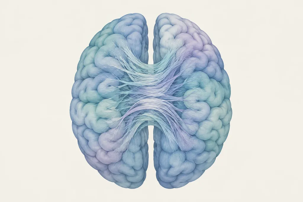
In MS, a brain scan can show changes in the tissue connecting the brain’s halves, but it cannot confirm that nerve signals have failed.
What They Found
This opinion paper examines how brain scans are used to estimate damage to connections between the brain’s halves. Such scans can measure tissue loss, but they cannot show whether nerve signals still travel through that tissue. The authors discuss an earlier study that found a link between brain structure and function in people without MS, but not in people with early relapsing MS. They say claims about signal failure need tests of function that match the brain area and task being studied. A scan finding used to predict future outcomes needs a different check: whether its predictions hold up in other groups.
Who Should Care and Why
This paper discusses people with early relapsing MS and people without MS from an earlier study. It does not report a new study of people with MS. It is about how researchers interpret brain scans, especially when studying thinking problems in MS. The distinction matters because tissue loss and failed nerve signaling are not the same finding. The authors also separate a scan’s ability to predict an outcome from its value in care decisions.
Important Considerations
You should know this is an opinion paper, not a new test of patients or a treatment. The abstract does not give the size of the earlier study it discusses. It does not show whether any scan finding improves decisions about your care.
AI-generated summary — for informational purposes only, not medical advice
Read the original abstract
Published in Journal of clinical neuroscience : official journal of the Neurosurgical Society of Australasia by . Reproduced from the PubMed record.
Automated magnetic resonance imaging can quantify callosal atrophy and estimate structural disconnection in multiple sclerosis, but neither measurement establishes physiological conduction failure. In an early relapsing-remitting cohort, Wahl and colleagues found a relationship between motor-callosal microstructure and interhemispheric inhibition in controls that was not detectable in patients. This finding illustrates why a structural feature cannot automatically inherit a functional interpretation. Earlier structure-function studies provide a foundation for contemporary pipelines when the measurements, pathways, and populations are comparable. This opinion paper organizes validation into five evidence domains with separate biological and prognostic branches: technical validity, biological validity, physiological validity, prognostic performance, and clinical utility. Physiological claims require functional evidence matched to anatomy and task, with reliability and competing circuit explanations assessed. A purely prognostic feature instead requires credible predictive evaluation, including external validation and calibration; it may be useful as a proxy for global disease burden. Clinical utility concerns the consequences of a specified decision and is distinct from predictive performance and implementation feasibility. Reports should preserve the qualifier in estimated structural disconnection and reserve physiological interpretations for measurements supported by corresponding functional evidence.
Categories
Explore more research
Check out the MS Buddy app
It’s free, with no ads. Built by people living with MS.


Related MS research
11/18/2026
An adjusted MRI scan found more veins within MS brain lesions in 17 people with MS. It improved scan
Read More
1/1/2026
After a car crash, brain and spine scans revealed signs of MS in an 18-year-old male. Further tests
Read More
11/15/2026
In 70 people with MS in Brazil, blood tests often showed signs of past viral exposure. Results did n
Read More
11/12/2026
In a study of 601 people in Japan after first optic nerve inflammation, some later had MS. Others ha
Read More
11/12/2026
A new way to analyze brain scans generally predicted thinking test scores in people with MS better t
Read More
11/11/2026
Brain scans of people with low-disability MS showed subtle differences from scans of people without
Read MoreNot all studies carry the same weight: sample size, study design and whether results were replicated all matter. The credibility score above reflects the journal, and the original paper is linked under More Details. Read how MS Buddy selects and scores research, and talk to your MS care team before acting on any study.