MS tissue and mice show damage in a brain vision hub
By MS Buddy ·
Summarized from Acta neuropathologica, Nov 2026 · PubMed. AI-generated summary. Not medical advice.
Animal study · donated brain tissue from people with MS and chemically exposed mice
Research by Jackson David McGrath, Sebastian Werneburg and 3 colleagues at the University of Michigan in the United States.
What damage occurs in a less-studied brain vision area in MS and mice?
Key Takeaway
Donated MS brain tissue showed nerve covering loss in a vision hub, while mice showed sex differences in related immune cell activity.
What They Found
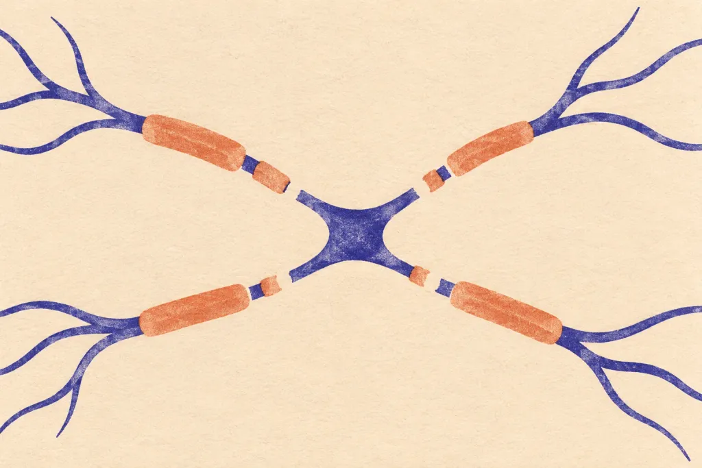
Donated MS brain tissue showed loss of myelin (the protective covering around nerves) in a brain area that helps process vision. Mice exposed to a chemical that damages nerve covering developed vision problems, without detectable loss of nerve cells or damage to nerve fibers. Male mice developed a distinct area of increased immune cell activity, where more connections between nerve cells were removed. After chemical exposure stopped, vision and nerve connections partly recovered, but a core area of increased immune cell activity remained. Female mice also developed widespread nerve covering loss and increased immune cell activity, but without the same distinct pattern.
Who Should Care and Why
This study examined donated brain tissue from people with MS and mice exposed to a chemical that damages nerve covering. The human findings identify damage in a brain area that helps process vision. This area has been less clearly understood than other parts of the visual system in MS. The mouse findings link increased immune cell activity with loss of connections between nerve cells. The differences between male and female mice provide a way to study how sex affects these patterns.
Important Considerations
The study combined donated human brain tissue analysis with mouse experiments, but the abstract gives no sample sizes. The findings about vision recovery and sex differences came from mice, not people with MS. This study does not show whether you would experience the same patterns or recovery.
AI-generated summary — for informational purposes only, not medical advice
Read the original abstract
Published in Acta neuropathologica by et al.. Reproduced from the PubMed record.
Visual impairment is a common and clinically salient manifestation of Multiple Sclerosis (MS), yet pathology across visual system structures remains incompletely defined. Although the optic nerve, lateral geniculate nucleus, and visual cortex have been extensively studied, involvement of the superior colliculus (SC), a key visual processing hub, remains unclear. Here, we combined human postmortem tissue analysis with functional assessment and spatial mapping in the MS-relevant cuprizone (CPZ) mouse model to define how demyelination and secondary injury are organized within the SC. Postmortem SC tissue from donors with MS revealed myelin loss, including focal demyelinated lesions. In mice, CPZ impaired visual function and induced widespread SC demyelination without detectable neuronal loss or axonal degeneration. In male mice, although diffuse demyelination was accompanied by widespread microgliosis, atlas-based mapping uncovered a previously unrecognized spatial organization: a discrete high-microgliosis compartment with a stereotyped location and shape in every CPZ-treated mouse. This compartment did not correspond to canonical SC maps and was not explained by baseline microglial or myelin differences or by variability in local demyelination severity. Instead, high-microgliosis regions showed increased synaptic elimination, suggesting that secondary synaptic pathology may contribute to spatial organization of microgliosis beyond diffuse myelin loss alone. Prolonged CPZ exposure expanded the high-microgliosis compartment stereotypically, whereas withdrawal produced spatially ordered partial resolution with persistence of a high-microgliosis core and partial visual recovery. Former high-microgliosis territories showed partial restoration of synaptic density despite myelin recovery comparable to persistent high-microgliosis regions. In contrast, female CPZ-treated mice developed widespread SC demyelination and microgliosis without comparable compartmentalization, demonstrating sex-differential organization of microglial reactivity. Together, these findings identify the human SC as a site of demyelinating injury in MS and establish the CPZ-treated mouse SC as a sex-sensitive in vivo framework for investigating spatially patterned microglial reactivity, synaptic pathology, and incomplete resolution of microgliosis after demyelination.
Categories
Explore more research
Check out the MS Buddy app
It’s free, with no ads. Built by people living with MS.


Related MS research
11/17/2026
An experimental drug eased nerve-related problems and reduced inflammation in mice with an MS-like d
Read More
11/17/2026
A review explores iron-driven cell death in MS and other diseases involving immune attacks on the bo
Read More
11/16/2026
In mice with damaged nerve coverings, fat-derived stem cells treated with metformin improved repair.
Read More
11/17/2026
A review suggests MS fatigue may reflect extra effort to control movement and incomplete recovery af
Read More
11/16/2026
Computer studies found MS fatigue surveys can give similar group results but different individual sc
Read More
11/17/2026
A review found exercise, therapy focused on thoughts and behavior, and self-management programs show
Read MoreNot all studies carry the same weight: sample size, study design and whether results were replicated all matter. The credibility score above reflects the journal, and the original paper is linked under More Details. Read how MS Buddy selects and scores research, and talk to your MS care team before acting on any study.