MS brain scans found changes that failed a stricter test
By MS Buddy ·
Summarized from Journal of magnetic resonance imaging : JMRI, Nov 2026 · PubMed · DOI. AI-generated summary. Not medical advice.
Observational study · 40 people with low-disability MS and people without MS
Research by Fabiana Bianca Hayashi Umezaki, Elisa Harumi Kozasa and 7 colleagues at Hospital Israelita Albert Einstein in Brazil.
Do brain scans show changes in people with MS who have low disability?
Key Takeaway
Brain scans showed subtle differences in people with MS and low disability, but those findings did not hold up under a stricter analysis.
What They Found
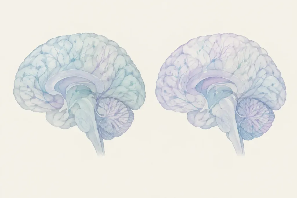
Researchers compared brain scans from people with MS and people without MS. Everyone with MS could walk without help. An initial analysis found differences in several areas of white matter, which carries messages through the brain. It also found differences in white matter that looked normal on standard scans. None of these differences remained clear when researchers used a stricter analysis.
Who Should Care and Why
This study included people with MS and people without MS. The people with MS had low levels of disability and could walk without help. The study asked whether scans could detect changes even when parts of the brain looked normal. Its findings may interest people trying to understand what brain scans show in low-disability MS. The stricter analysis leaves uncertainty about whether the reported differences were reliable.
Important Considerations
You should know this study compared scans from 20 people with MS and 20 people without MS at one point in time. It did not track whether scan differences predicted later disability. You cannot tell from this study whether the initial findings would hold up in a larger group.
AI-generated summary — for informational purposes only, not medical advice
Read the original abstract
Published in Journal of magnetic resonance imaging : JMRI by et al.. Reproduced from the PubMed record.
Diffusion tensor imaging (DTI) is sensitive to white matter (WM) microstructural alterations in multiple sclerosis (MS), but duration-defined "early MS" cohorts may be clinically heterogeneous. To investigate diffusion alterations in WM and normal-appearing white matter (NAWM) in low-disability MS versus healthy controls (HC). Retrospective cross-sectional case-control study. 20 patients with MS (Expanded Disability Status Scale ≤ 3.5; ambulatory without assistance) and 20 age- and sex-matched HC. 3.0 T; 3D T1-weighted and FLAIR sequences and a single-shot spin-echo echo-planar sequence with 60 diffusion directions (b = 1000 s/mm2) and nine b0 volumes. Tract-Based Spatial Statistics (TBSS) assessed fractional anisotropy (FA), axial diffusivity (AD), radial diffusivity (RD), and mean diffusivity (MD) in conventional WM and lesion-masked NAWM. DeepLesionBrain (volBrain) automatically segmented WM lesions; a neuroradiologist with 10 years of experience reviewed and corrected segmentations when necessary. FMRIB Software Library (FSL) randomize using 10,000 permutations, threshold-free cluster enhancement, and family-wise error correction (p < 0.01). Permutation Analysis of Linear Models (PALM) used two-sided inference and FWE correction across diffusion metrics and directions. Standard TBSS showed lower FA in the right anterior corona radiata (ACR) and posterior thalamic radiation (PTR), and higher RD in the corpus callosum genu, in MS versus HC. NAWM-restricted TBSS showed lower FA in the corpus callosum genu, right PTR, and right tapetum, and higher RD in bilateral ACR, right posterior corona radiata, and an unclassified region. In randomized, AD and MD were nonsignificant (minimum corrected p = 0.0118 and 0.0103 in standard TBSS; p = 0.0103 and 0.0107 in NAWM, respectively). Under PALM across-metric correction, no metric remained significant: standard TBSS, FA p = 0.0861, AD p = 0.1273, RD p = 0.0271, MD p = 0.0177; NAWM, FA p = 0.0807, AD p = 0.1225, RD p = 0.0282, MD p = 0.0197. Functionally defined low-disability MS shows focal WM and NAWM microstructural alterations, predominantly reflected by reduced FA and increased RD. 4. Stage 1.
Categories
Explore more research
Check out the MS Buddy app
It’s free, with no ads. Built by people living with MS.


Related MS research
11/17/2026
An experimental drug eased nerve-related problems and reduced inflammation in mice with an MS-like d
Read More
11/17/2026
A review explores iron-driven cell death in MS and other diseases involving immune attacks on the bo
Read More
11/18/2026
An adjusted MRI scan found more veins within MS brain lesions in 17 people with MS. It improved scan
Read More
11/16/2026
In mice with damaged nerve coverings, fat-derived stem cells treated with metformin improved repair.
Read More
11/17/2026
Genetic and brain scan data linked inherited brain structure patterns to MS risk. Researchers also f
Read More
11/17/2026
Donated MS brain tissue showed damage in a vision hub. Mouse findings linked local immune cell activ
Read MoreNot all studies carry the same weight: sample size, study design and whether results were replicated all matter. The credibility score above reflects the journal, and the original paper is linked under More Details. Read how MS Buddy selects and scores research, and talk to your MS care team before acting on any study.